Left Atrium
Emergency Flail Mitral Leaflet: Acute Severe Mitral Regurgitation
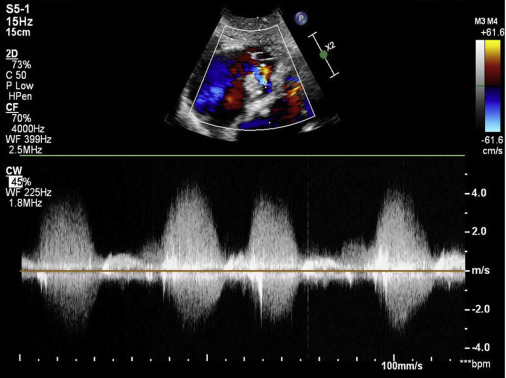
The patient presented with a history of chronic shortness of breath, prompting transthoracic echocardiographic assessment. Imaging identified a flail posterior mitral leaflet associated with severe eccentric mitral regurgitation. Doppler findings further suggested significant pulmonary hypertension, including a short pulmonary artery acceleration time and mid-systolic notching/"flying W" morphology of the RVOT/pulmonary flow profile.
Aortic Valve
Paediatric Rheumatic Heart Disease: Severe Multivalvular Regurgitation
An 8-year-old with known rheumatic heart disease was referred for surgical evaluation. Echocardiography demonstrated severe multivalvular involvement with a flail anterior mitral leaflet, rheumatic leaflet thickening/restriction, severe mitral regurgitation, severe aortic regurgitation, severe tricuspid regurgitation, and associated chamber dilatation.
Atria
Infiltrative Cardiomyopathy Pattern
A patient in their 70s recently diagnosed with hypertension presenting with progressive heart failure symptoms, including dyspnea, easy fatigability, bilateral lower-extremity edema, and paroxysmal nocturnal dyspnea. Echocardiographic findings demonstrated a pattern concerning for an infiltrative cardiomyopathy, including possible cardiac amyloidosis, prompting further diagnostic workup. An additional mass-like structure within the right atrioventricular groove was also identified and requires further characterization.
Aortic Root
Aortic Root and Ascending Aortic Aneurysm
An elderly patient with a known aortic root and ascending aortic aneurysm, diagnosed approximately two years earlier, underwent transthoracic echocardiography for interval surveillance. The study focused on serial aortic dimensions, aortic valve morphology and function, presence of aortic regurgitation, left ventricular size and systolic performance, and any evidence of hemodynamic impact related to the aneurysm.
Aortic Valve
Subaortic Membrane: Subvalvular LVOT Obstruction
A young adult male with longstanding intermittent palpitations and previously preserved tolerance of routine activities presented only after progressive symptoms culminated in a near-syncopal episode with profuse diaphoresis.
Aorta
Tetralogy of Fallot In A Middle-Aged Adult
A 55-year-old lifelong farmer with previously unrestricted functional capacity presented with a 2-month history of progressive exertional dyspnea. Echocardiography demonstrated a large perimembranous VSD with bidirectional flow, severe valvular pulmonary stenosis, overriding aorta, elevated RV systolic pressure, and an echogenic pulmonary valve mass under further evaluation.
IVC
Tuberculous Pericarditis: Effusion and Constrictive Physiology
Male in early-20s with a prior history of pulmonary tuberculosis referred for echo after chest radiography demonstrated a water-bottle cardiac silhouette. Patient presented with pleuritic/positional chest pain, orthopnea, dyspnea, palpitations, bilateral lower-extremity edema, weight loss, fever, and marked resting hypoxemia.
Aortic Root
Sinus of Valsalva Aneurysm: RCC Rupture Into the LVOT
A rare ruptured right coronary sinus of Valsalva aneurysm extending into the LVOT, producing abnormal aorta-to-left ventricular flow with potential associated aortic regurgitation and LVOT hemodynamic disturbance.
Atrial Septum
Embolic-Stroke: Pulmonary Arteriovenous Malformation
A contrast-echocardiography pitfall in which rapid left-heart bubbles initially resemble an intracardiac shunt, while careful localization and CT angiography identify a pulmonary arteriovenous malformation.
Left Atrium
NBTE: Severe Mitral Stenosis, and Mechanical Valve Thrombosis
A Doppler-focused case following antiphospholipid-syndrome–associated nonbacterial thrombotic endocarditis from severe native mitral obstruction through mechanical mitral valve thrombosis.
Left Ventricle
Incidental Left Ventricular Pseudoaneurysm
An incidentally detected inferolateral left-ventricular outpouching in an older adult, with echocardiography and multimodality imaging used to distinguish chronic pseudoaneurysm from other ventricular outpouchings.
Left Atrium
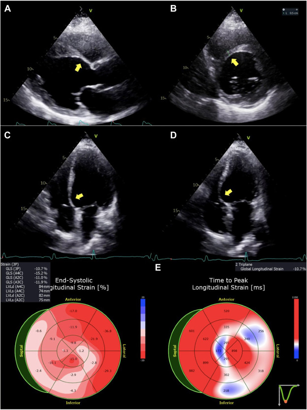
Cor Triatriatum Sinister With Symptomatic Left-Atrial Inflow Obstruction
An adult presentation of cor triatriatum sinister in which multimodality imaging and invasive hemodynamics define a restrictive left-atrial membrane before surgical resection.
 0 views
Interventricular Septum
Cardiac Sarcoidosis Associated With IBD
An inflammatory cardiomyopathy case linking systemic disease, conduction abnormalities, ventricular dysfunction, PET/CMR findings, and arrhythmic risk.
Left Ventricular Apex
Blueberry-on-Top Phenomenon in Apical HCM
An apical-HCM case focused on a small residual apical cavity, severe apical hypertrophy, cavity obliteration, aneurysm/thrombus search, and contrast/CMR confirmation.
Mitral Valve
Contrast Echo Revealing Eccentric Mitral Regurgitation
A case showing how ultrasound-enhancing agents and improved endocardial definition can reveal the mechanism or true extent of an eccentric MR jet.
Right Ventricle
Hypoplastic TV Leaflet and Right-Heart Failure
A congenital valvular case highlighting tricuspid leaflet hypoplasia, malcoaptation, and severe TR.
Aortic Valve
Aortic Regurgitation Defined by 3D Echo
A mechanism-focused aortic-regurgitation case using 3D echocardiography to identify subtle cusp pathology that is difficult to appreciate on standard 2D views.
Left Ventricular Outflow Tract
Mitral Regurgitation and Dynamic LVOT Obstruction: Hemodynamic Interplay
A physiology case showing how systolic anterior motion, LVOT obstruction, and mitral regurgitation influence each other and complicate Doppler interpretation.
Pulmonary Artery
Saddle Pulmonary Embolism Seen on Transthoracic Echocardiography
An acute pulmonary-embolism case in which thrombus is visualized at the pulmonary-artery bifurcation, with complete RV pressure-overload assessment.
Pulmonary Valve
Tricuspid Atresia With Absent Pulmonary Valve Syndrome
A complex congenital case combining tricuspid atresia and absent pulmonary valve physiology, with emphasis on segmental anatomy and pulmonary artery dilation.
Cardiac Chambers
A Rapidly Growing Cardiac Mass: Malignant or Benign?
A diagnostic-reasoning case for a fast-changing intracardiac mass, integrating serial echocardiography, attachment, perfusion, CMR, and pathology.
Mitral Valve
Papillary Muscle Lipoma in an Adolescent
A benign cardiac-mass case focused on a papillary-muscle lipoma and differentiation from thrombus, fibroma, vegetation, and normal variants.
Aortic Root
Bioprosthetic Aortic Valve Dehiscence and Acute Regurgitation
A prosthetic-valve emergency case focused on abnormal rocking motion, dehiscence, paravalvular regurgitation, endocarditis, and cardiogenic shock.
Left Atrial Appendage
Giant Left Atrial Thrombus
A mass-differential case focused on a very large left-atrial thrombus, mobility, attachment, valve obstruction, embolic risk, and distinction from tumor.
Right Ventricle
Catastrophic Bone Cement Implantation Syndrome
A perioperative shock case illustrating sudden RV failure, pulmonary vascular obstruction, and the role of transesophageal echocardiography during bone cement implantation syndrome.
Congenital
Atypical Venous Drainage, R-SVC & Dilated Coronary Sinus
An atypical systemic venous drainage pattern featuring a right superior vena cava (R-SVC) with a markedly dilated coronary sinus, prompting evaluation for associated anomalous venous return, persistent left-sided SVC, or other congenital systemic venous variants.
Coronary Sinus
Caval and Coronary Sinus Venous Anomalies: Four-Pattern Review
A multi-case for persistent left superior vena cava and related systemic venous anomalies, integrating TTE, agitated saline, CT, and procedural relevance.
Left Ventricular Outflow Tract
Acquired Dynamic Left Ventricular Outflow Tract Obstruction
A hemodynamic case on dynamic LVOT obstruction outside classic hypertrophic cardiomyopathy, integrating loading conditions, SAM, Doppler morphology, and treatment response.
Aortic Root
Congenital Left Main Coronary Atresia
A congenital coronary anomaly case emphasizing indirect echocardiographic clues, collateral flow, ventricular dysfunction, and confirmatory CT or angiography.
Left Ventricular Apex
Post-COVID Myocarditis With Large Apical LV Thrombus
A case combining myocarditis, reduced LV function, and a large apical thrombus discovered during cardiac screening after COVID-19.
 1 view
Mitral Annulus
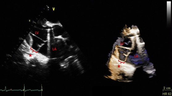
Mitral Annular Disjunction in Marfan Syndrome
A case focused on recognizing mitral annular disjunction, associated prolapse and regurgitation, and arrhythmic-risk imaging features in Marfan syndrome.
Inferior Vena Cava
Hepatic Cyst–Induced Internal Jugular Venous Thrombosis
A case linking a large hepatic cyst, right-sided venous compression, and internal jugular thrombosis, with focused ultrasound and hemodynamic assessment.
 6 views
Aortic Valve
Acquired Gerbode Defect After Aortic Valve Endocarditis
A case on an acquired left-ventricle-to-right-atrium shunt caused by infective endocarditis, highlighting color Doppler localization and differentiation from tricuspid regurgitation.
Pericardium
Granulomatosis With Polyangiitis With Pericarditis
A multimodality case examining pericardial inflammation and effusion in granulomatosis with polyangiitis, with emphasis on echo findings and CMR correlation.
No cases match the current filters.