Transthoracic echocardiogram with circumferential pericardial effusion on parasternal short-axis view.
Transthoracic echocardiogram with circumferential pericardial effusion on apical four-chamber view.
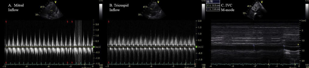
Pulsed-wave-Doppler recordings of (A) mitral and (B) tricuspid inflow showing mild respiratory variation of mitral and tricuspid inflows in setting of moderate pericardial effusion. (C) M-mode echocardiogram of IVC showing dilated IVC (>21 mm diameter) with plethora.
On-treatment TTE videos of patient 1 showing no pericardial effusion or signs of pericardial constriction on parasternal short-axis view.
On-treatment TTE videos of patient 1 showing no pericardial effusion or signs of pericardial constriction on parasternal four-chamber view.
CCBY_001
Granulomatosis With Polyangiitis With Pericarditis
clinical_presentation
A patient with established or suspected systemic vasculitis develops chest discomfort, dyspnea, inflammatory symptoms, or new electrocardiographic changes. Echocardiography is requested to evaluate pericardial disease and ventricular function.
imaging_context
- modality
- Transthoracic Echocardiogram
- primary_view
- PSSAX effusion loop; Ap4_Ch loop; Doppler
- media_type
- Echo Cineloops, 2D Still Doppler Image
key_findings
Pericardial thickening or effusion may be present.
Assess for tamponade physiology rather than relying on effusion size alone.
Global and regional ventricular function should be documented.
CMR can add tissue-characterization evidence of active inflammation.
measurements
| parameter | value | reference / interpretation |
|---|---|---|
| Effusion dimensions by location | 1.8 cm | Record maximal end-diastolic separation by location. Hemodynamic significance depends on chamber collapse, respiratory Doppler variation, venous/RAP findings, rate of accumulation, and clinical status—not size alone. |
| IVC size and collapsibility | >21 mm, <50% | Use IVC diameter and inspiratory change only in the appropriate spontaneous-breathing context. RAP estimation is less reliable with positive-pressure ventilation, abnormal respiratory effort, or unusual venous compliance. |
| Respiratory inflow variation | Mild | Measure respiratory variation from consistent Doppler sample positions and interpret with breathing pattern, rhythm, ventilation mode, and other echocardiographic signs of tamponade physiology. |
discussion
learning_points
4 points- Systemic vasculitis can involve the pericardium even when pulmonary or renal findings dominate.
- Effusion size does not determine tamponade; integrate chamber collapse, respiratory variation, IVC findings, and clinical status.
- Document ventricular function and regional wall motion to search for myocardial or ischemic involvement.
- Use multimodality imaging when echo findings and the clinical picture are discordant.
case_documentationReferences & attribution2 items+
references
- Ahmed T, Meredith D, Klein AL. Granulomatosis With Polyangiitis (Wegener's Granulomatosis) Complicated by Pericarditis: Our Experience of Two Cases. CASE: Cardiovascular Imaging Case Reports (2021). DOI: 10.1016/j.case.2020.11.008. Licensed under CC BY 4.0.
media_and_source_credits
- CASE JOURNALCC BY 4.0
Granulomatosis With Polyangiitis (Wegener's Granulomatosis) Complicated by Pericarditis: Our Experience of Two Cases
Ahmed T, Meredith D, Klein AL. · CASE: Cardiovascular Imaging Case Reports · DOI 10.1016/j.case.2020.11.008
Changes: Adapted for ECHOLOGISTS educational presentation; explanatory structure and formatting modified.